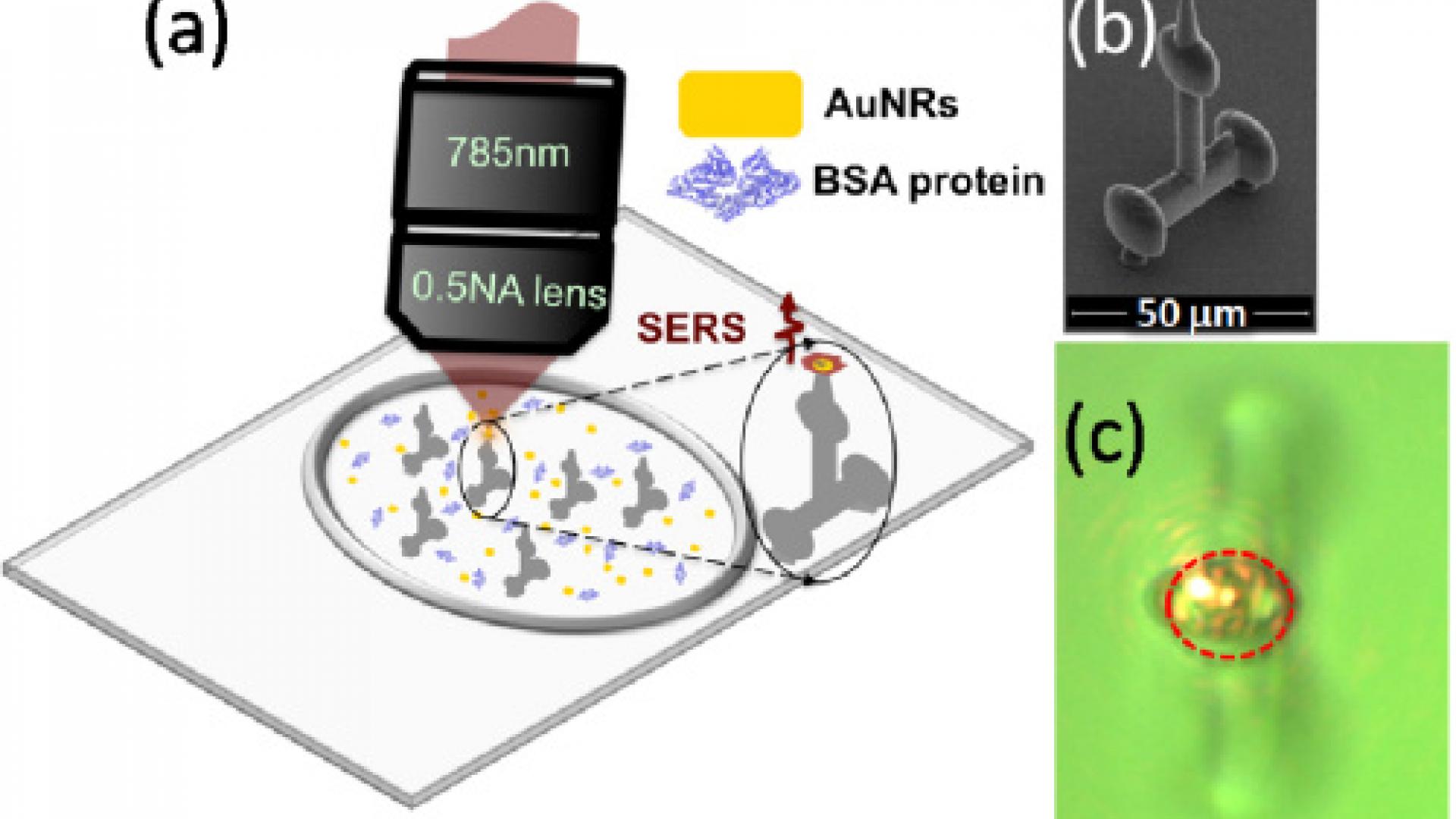
Fig. 1. (a) Sketch of the experimental setup. AuNRs dispersed in water are mixed and stabilized with BSA protein molecules [23]. The sample is placed in a micro-cell where TPP microstructures (tripods) are present. Upon illumination with a focused laser beam, the radiation pressure pushes the BSA-AuNR complexes within the focal spot, creating SERS-active aggregates and controlled decoration of the tripod tip in few minutes. The aggregation dynamics is monitored by detecting the BSA SERS spectrum at different times. (b) SEM image of one of the tripods studied in this Letter. (c) Microscope image of a tripod tip, with the laser beam focused on it (red dashed ring).
Optical forces are used to push and aggregate gold nanorods onto several substrates creating surface-enhanced Raman scattering (SERS) active hot spots for Raman-based identification of proteins. By monitoring the increase of the protein SERS signal, we observe different aggregation times for different curvatures of the substrates. The slower aggregation dynamics on curved surfaces is justified by a simple geometrical model. In particular, this technique is used to decorate three-dimensional microstructures and to quickly realize hybrid micro/nanosensors for highly sensitive detection of biological material directly in a liquid environment.
https://www.osapublishing.org/ol/abstract.cfm?uri=ol-43-20-5170
Related Persons

Assistant Professor,
Bioscience