Smarter MRI image analysis for the whole heart
A Bayesian approach that selects the most informative frames for manual labeling could speed up real-time heart MRI analysis.
About
Cardiovascular diseases remain a leading cause of death worldwide. Every year thousands of children are born with congenital heart disease. Reliable, efficient tools that can accurately assess heart function are critical for heart disease diagnosis and treatments.
Now, a novel machine learning method of analyzing real-time cardiac magnetic resonance imaging (MRI) scans has been developed by KAUST researcher Raúl Tempone in collaboration with scientists at the German Aerospace Center, in Cologne[1].
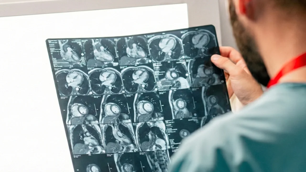
“Cardiac real-time MRI is an invaluable method of scanning the heart, and can acquire up to 50 frames per second, which is excellent in clinical terms,” says Tempone. “However, this generates thousands of images that are near-impossible to process manually.”
A typical short scan at 30 frames per second for 10 seconds can produce around 4,500 images to annotate across 15 ‘slices’, or cross-sectional images through the heart. Neural networks can accurately segment most of these images, but fail to consistently segment the ventricular cavity in the outer slices at the base and apex of the heart. This means that the ventricular volume — the amount of blood inside the heart’s ventricles throughout the cardiac cycle — in the outer slices is not always estimated reliably by the neural networks.
Read the full story on KAUST Discovery.