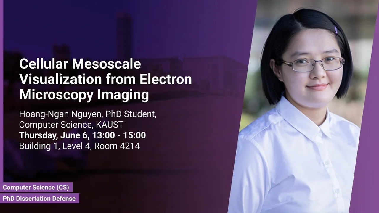
Cellular Mesoscale Visualization from Electron Microscopy Imaging
Currently, the acquisition of accurate cryogenic electron microscopy data deals with problems with complex and time-consuming processes, low signal-to-noise ratio, and missing wedge, leading to a lack of highly accurate imaging data. Such data would be necessary to develop computational methods/visualizations and essential to train deep learning models that are used to solve inverse problems.
Overview
Abstract
Currently, the acquisition of accurate cryogenic electron microscopy data deals with problems with complex and time-consuming processes, low signal-to-noise ratio, and missing wedge, leading to a lack of highly accurate imaging data. Such data would be necessary to develop computational methods/visualizations and essential to train deep learning models that are used to solve inverse problems.
In this research, we develop a system that generates synthetic data indistinguishable from the actual data. The proposed system also generates the data in a differentiable manner, allowing for gradient-based optimization for parameters of the electron microscope, denoising, and reconstruction. It contains three main components. First, a new rapid modeling tool allows users to model mesoscale biological structures with a few efforts. Second, a differentiable simulator receives modeled mesoscale biological structures, generates synthetic data, and supports learning. Third, a visualization tool gives high-quality visualization of cryogenic electron microscopy data and can be used as an evaluation tool to assess synthetic data. We evaluate the quality of the synthetic data, the correctness of interpretation from the learning model, visualization with the real-world specimen data, and the support from bio-experts.
Brief Biography
Ngan Nguyen is a Ph.D. student at NANOVIS group under the supervision of Professor Ivan Viola. Her research focuses on computer graphics, visualization and their overlap with animation and biomechanics.

