A new computer-aided diagnostic tool developed by KAUST scientists could help overcome some of the challenges of monitoring lung health following viral infection.
Like other respiratory illnesses, COVID-19 can cause lasting harm to the lungs, but doctors have struggled to visualize this damage. Conventional chest scans do not reliably detect signs of lung scarring and other pulmonary abnormalities, which makes it difficult to track the health and recovery of people with persistent breathing problems and other post-COVID complications.
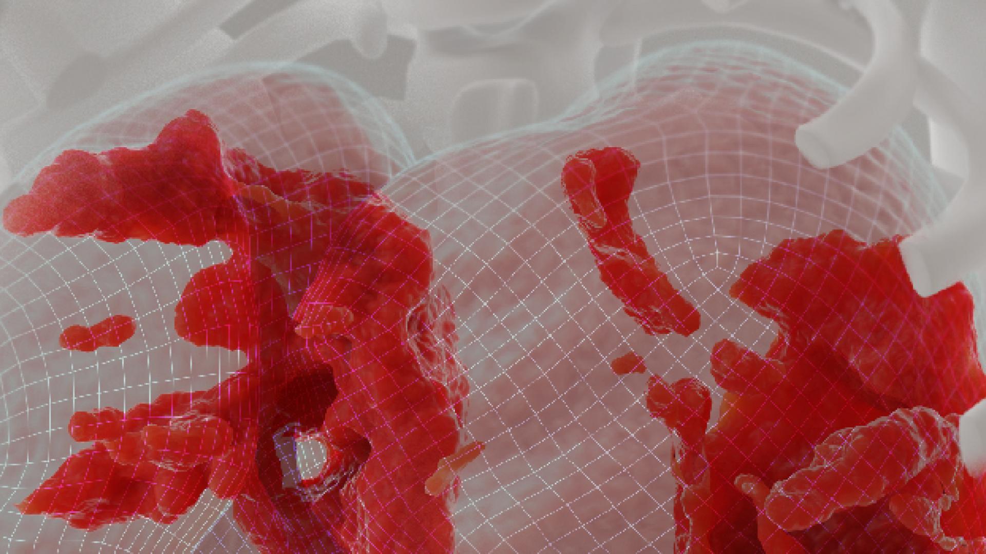
The new method developed by KAUST — known as Deep-Lung Parenchyma-Enhancing (DLPE) — overlays artificial intelligence algorithms on top of standard chest imaging data to reveal otherwise indiscernible visual features indicative of lung dysfunction.
Through DLPE augmentation, “radiologists can discover and analyze novel sub-visual lung lesions,” says computer scientist and computational biologist Xin Gao. “Analysis of these lesions could then help explain patients’ respiratory symptoms,” allowing for better disease management and treatment, he adds.
Gao and members of his Structural and Functional Bioinformatics Group and the Computational Bioscience Research Center created the tool, along with artificial intelligence researcher and current KAUST Provost Lawrence Carin and clinical collaborators from Harbin Medical University in China.
The method first eliminates any anatomical features not associated with the lung parenchyma; the tissues involved in gas exchange serve as the main sites of COVID-19–induced damage. That means removing airways and blood vessels, and then enhancing the pictures of what is left behind to expose lesions that might be missed without the computer’s help.
The researchers trained and validated their algorithms using computed tomography (CT) chest scans from thousands of people hospitalized with COVID-19 in China. They refined the method with input from expert radiologists and then applied DLPE in a prospective fashion for dozens of COVID-19 survivors with lung problems, all of whom had experienced severe disease requiring intensive care treatment.
In this way, Gao and his colleagues demonstrated that the tool could reveal signs of pulmonary fibrosis in COVID long-haulers, thus helping to account for shortness of breath, coughing and other lung troubles. A diagnosis, he suggests, that would be impossible with standard CT image analytics.
Read the full text.