© 2020 KAUST
Living cells inside the body could be placed under surveillance—their location and migration noninvasively tracked in real time over many days—using a new method developed by researchers at KAUST.
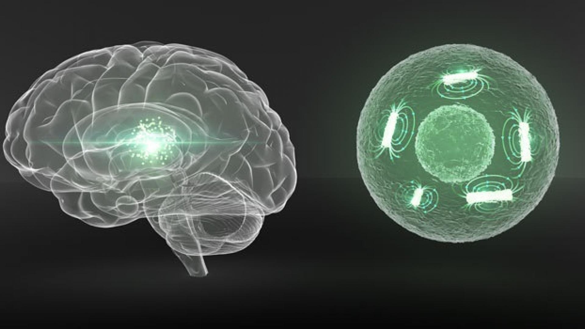
The technique uses magnetic core-shell iron nanowires as nontoxic contrast agents, which can be implanted into live cells, lighting up those cells’ location inside a living organism when scanned by magnetic resonance imaging (MRI). The technique could have applications ranging from studying and treating cancer to tracking live-cell medical treatments, such as stem cell therapies.
Jürgen Kosel and his team recently showed that core-shell iron nanowires could selectively kill cancer cells with a combination attack, delivering an anticancer drug into target cells while also puncturing the cell’s membrane and unleashing blasts of heat. Now, in collaboration with researchers from the CIC biomaGUNE in San Sebastian, Spain, the team has shown that the same type of iron core, iron-oxide shell nanowires, can be used for noninvasive medical imaging. The nanowires could potentially be used as "theranostic" agents, able to identify, track and then take out target cells.
“Cell labeling and tracking has become an invaluable tool for scientific and clinical applications,” says Aldo Martínez-Banderas, a Ph.D. student in Kosel’s team. “One of the key aspects of cell tracking studies is the sensitivity to detect a small number of cells after implantation, so the strong magnetization and biocompatibility of our nanowires are advantageous characteristics for MRI tracking.”
Read the full article
Related Persons