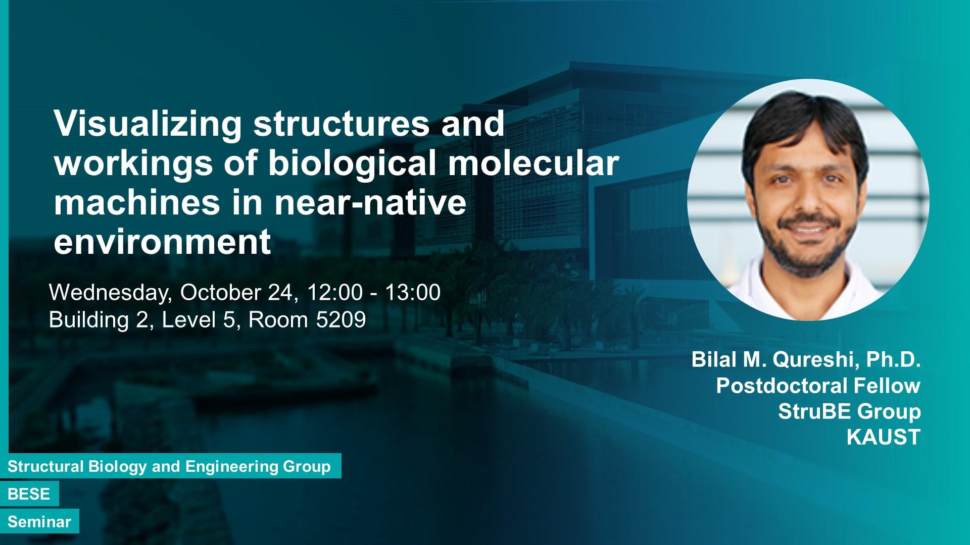
Structural insights into biological molecules such as proteins at atomic detail help us understand their properties, functions, interactions and regulation. This allows us to understand biological processes at the molecular and cellular level. Cryo electron microscopy of vitrified biological macromolecular complexes allows obtaining structural insight in near-native conditions. Moreover, the method is most suitable for studying macromolecular complexes with multiple interacting subunits in multiple conformations. Thus besides resolving structures of individual components (subunits), we also learn about the way the molecular machinery is built and how it works. Some examples from ongoing projects in Arold lab e.g. Cdc48A – the plant unfoldase/degradation machinery will illustrate the power of cryo electron microscopy for studying these molecular machines.
Bio:
Dr. Bilal Qureshi I am a biophysicist by training and I investigate fundamental processes of life at molecular level. I employ methods like x-ray diffraction or single-particle cryo electron microscopy to obtain structures of biological macromolecules. Moreover, I investigate interactions of these molecules by biophysical and biochemical methods to understand the underlying molecular mechanisms for basic cellular processes such as signal-transduction, protein trafficking and enzyme regulation etc. Besides, I produce engineered molecules with predicted properties to further corroborate my findings. My research has impact in basic sciences as well as for drug development.
Professor,
Bioscience

