Source: KAUST Discovery article
Ultrashort peptides that spontaneously form intricate scaffolds are helping scientists understand how cells respond and adapt to three-dimensional environments that closely resemble the natural settings of our bodies.
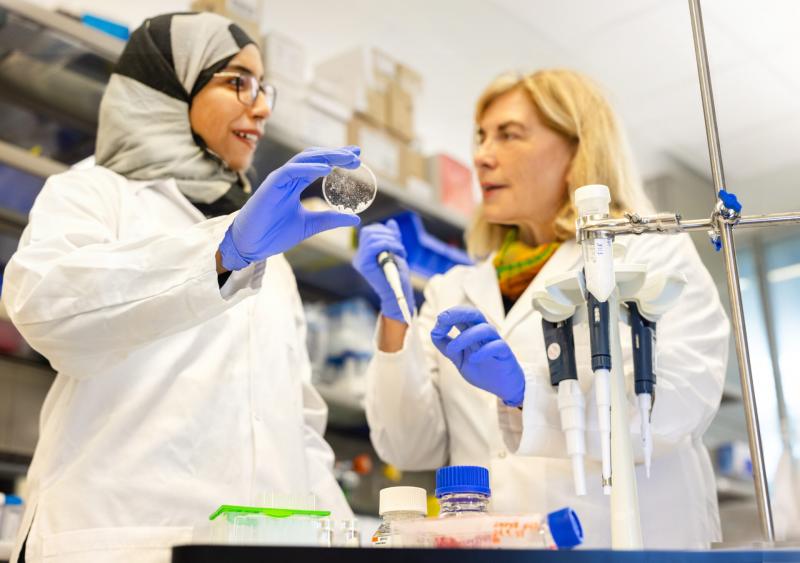
Developed by bioengineer Charlotte Hauser and her colleagues at KAUST, these self-assembling peptides give rise to a jelly-like framework that mimics the shape and mechanical properties of the extracellular matrix, the foundational support structure for cells.
Such surroundings in turn affect the genetics and metabolism of cultured cells, as Hauser and her team demonstrate.
The researchers analyzed human skin cells grown both in their 3D hydrogel and under conventional lab Petri dish conditions. In total, they observed differences in 1,833 genes and 29 metabolic pathways implicated variously in nutrient uptake, energy generation and other processes essential for proper cell functioning and health — a finding that underscores the importance of experimental culture conditions.
Those conditions are far from static. Notably, the KAUST team found that cells grown in their 3D hydrogel altered their environment, depositing proteins and physically interacting with the material in ways that changed the stiffness and composition of the complex scaffold. Similarly, the cells adapted and transformed in response to their surroundings, much like they would in the ever-changing milieu of the human body.
By emphasizing the dynamic interplay between cells and their microenvironment, these peptide-based scaffolds hold the promise of offering an enhanced platform for studying cellular behaviors, metabolism and gene expression,” says Sherin Abdelrahman, a postdoctoral fellow in Hauser’s research group and a co-first author of the new report.
According to Abdelrahman, the findings highlight the limitations of studying cells under simplified culture conditions. These traditional lab setups often fail to replicate the complexities of mechanical sensing and molecular biology, which could explain the frequent failures in developing drugs and regenerative therapies.
3D materials like the KAUST team’s peptide-based hydrogels hold promise to boost the success rate.
“Enhancing our understanding of the inherent characteristics of 3D matrices employed as scaffolds — particularly the utilization of ultrashort self-assembling peptides — offers the prospect of gaining better control over their properties.” Abdelrahman says. This allows for targeted modulation in alignment with specific research needs.
Computer simulations performed by co-first author Rui Ge, another postdoc in Hauser’s lab, demonstrated the assembly dynamics of these materials, drawing attention to the way that the packaging of different peptide fibers affects the stiffness of the overall scaffold.
Hauser now hopes to use this atomic-scale information, along with the cellular data, to optimize her group’s constructs for a range of biomedical applications. “There is an urgent demand for truly synthetic designer matrices for cell, tissue and organoid culturing,” she says.
REFERENCE
Abdelrahman, S., Ge, R., Susapto, H.H., Liu, Y., Samkari, F., Moretti, M., Liu, X., Hoehndorf, R., Emwas, A.-H., Jaremko, M., Rawas, R.H. & Hauser, C.A.E. The impact of mechanical cues on the metabolomic and transcriptomic profiles of human dermal fibroblasts cultured in ultrashort self-assembling peptide 3D scaffolds. ACS Nano 17, 14508–14531 (2023).| article