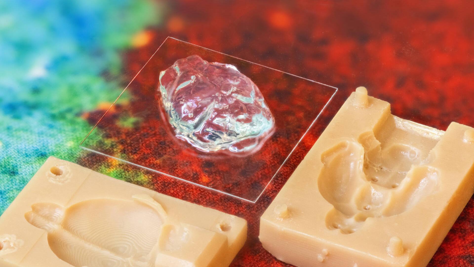
In a notable achievement at Charlotte Hauser’s laboratory in KAUST, postdoc Sherin Abdelrahman has established 3D models of brain organoids based on 3D scaffolds composed of ultrashort self-assembling peptides that contain nothing more than four amino acids.
These compounds are selected from a class of rationally designed ultrashort peptides, discovered by Hauser. Due to their unique sequences, the peptides are able to self-assemble into extracellular matrix-like scaffolds by mimicking the structure of collagen fibers. Abdelrahman incorporated dopaminergic as well as cortical neurons into the 3D peptide scaffold.
Before applying for a Ph.D. position with Hauser, Abdelrahman spent 10 years working at the nearby King Fahad Medical Research Center in Jeddah.
“Although my role at the Research Center gave me an excellent experience, I knew that there was more that I could explore with a Ph.D.” she recalls.
Initially, Hauser offered Abdelrahman an internship, and after only two weeks, she was accepted to the Ph.D. program. Abdelrahman remembers some of the challenges of adapting to academic life, but happily reports that she has developed a passion for neuroscience. “At some point, I would like to be able to call myself a neuroscientist,” she says.
“I enjoy research because it challenges my brain. I come to work every day not knowing what to expect, which is very exciting.”
After completing her Ph.D. in 2022, Abdelrahman is continuing her research on brain organoids, trying to develop a model for Parkinson’s disease using cutting-edge biomaterials and bioprinting technologies.
“The major goal of our research is to develop natural, but synthetic, sustainable biomaterials that serve as cellular matrices and mimic the native cellular microenvironment while preserving the functionality of the tissue,” says Hauser.
Promise for Parkinson’s
The key advantage of the novel peptides is that they self-assemble into hydrogel scaffolds under physiological conditions, without the need for harmful treatments, such as UV or chemical cross-linking that might affect cellular activity and proliferation.
Unlike 2D in-vitro cellular models, where the cells are cultured directly on plastic or glass surfaces, the peptide matrices are used to create 3D scaffolds where cells are embedded in a way that has a much closer resemblance to their native environment.
Hauser’s lab is at the forefront of 3D bioprinting research, applying innovative 3D peptide scaffolds as bioinks and developing a robotic printer for extremely accurate and consistent results.
“When we assessed the viability of the neurons inside our 3D peptide scaffolds, we found that the cells had better survival and proliferation inside our system compared with 2D controls,” says Abdelrahman.
“We were able to maintain the neuronal activity in our model for twice the time they were active in the 2D system.”
Their results showed that peptide scaffolds promoted dendritic branching, a morphological feature that is directly related to the formation of new synapses and thus has implications for the proper functioning of neurons.
“The interesting aspect of this finding is that some therapeutics used for the treatment of Parkinson’s disease induce increased branching,” says Hauser.
“We hope that our 3D model will provide a platform for dedicated studies of neurogenerative disorders, such as Parkinson’s disease, that may lead to more efficient treatment strategies,” she says, acknowledging the support of their collaborators in Saudi Arabia at Taif University and King Abdulaziz University.
As Abdelrahman notes, “When you want to recreate something that happens in-vivo, you need to first focus on recreating the main cell–cell and cell–microenvironment interactions that occur within the native tissue — in this case, the brain.
“In addition to the interactions between the cells and the microenvironment, we need to consider another vital aspect — the influence of the tissue geometry on cellular functionality. This aspect motivated us to print a mouse-brain-inspired model, accurately matching the real size of a mouse brain.
The significant differences observed between the 3D and 2D models undoubtedly have implications for drug testing, clinical trials and the development of new treatments.
This article has been sourced from the KAUST Insight website.